E3 binding protein also known as pyruvate dehydrogenase protein X component, mitochondrial is a protein that in humans is encoded by the PDHXgene.[5][6][7][8] The E3 binding protein is a component of the pyruvate dehydrogenase complex found only in eukaryotes. Defects in this gene are a cause of pyruvate dehydrogenase deficiency which results in neurological dysfunction and lactic acidosis in infancy and early childhood. This protein is also a minor antigen for antimitochondrial antibodies. These autoantibodies are present in nearly 95% of patients with primary biliary cholangitis, an autoimmune disease of the liver. In primary biliary cholangitis, activated T lymphocytes attack and destroy epithelial cells in the bile duct where this protein is abnormally distributed and overexpressed. Primary biliary cholangitis eventually leads to liver failure.[5]
Structure
The mRNA encoded by the human PDHX gene is approximately 2.5 kb in length, and expressed primarily in human skeletal and cardiac muscle tissues. The gene has been localized in humans to the 11th chromosome, with the specific location being 11p1.3.[9]
The protein encoded by the human PDHX gene, also known as E3 binding protein (E3BP), is part of the pyruvate dehydrogenase complex, a required complex for cellular respiration that catalyzes the dehydration of pyruvate to Acetyl-CoA.[10] The entire complex is 9.5 MDa in size, and has been described as 60-meric, meaning there are over 60 components that are assembled to make the entire complex. These subunits are conserved across many species, as the function of this complex is essential for the generation of ATP for all eukaryotes. The E3 binding protein directly interacts with the dihydrolipoamide transacetylase (E2) core, anchoring it to the complex. E3BP binds the I domain of E2 by its C-terminal I' domain. The composition of E2.E3BP was thought to be 60 E2 plus approximately 12 E3BP, however, equilibrium sedimentation and small angle x-ray scattering studies showed that the E3BP/E2 binding complex has a lower mass than the E2 subunit alone. Additionally, these studies showed that E3 binds to E2.E3BP outside the central dodecahedron of the PDH complex, and that this interaction creates a lower binding affinity for the E1 subunit. Together, these data support a substitution model, in which the smaller E3BP subunits replace the E2 subunits rather than adding to the 60-mer entire complex. The specific model illustrates 12 I domains of E2 being substituted by 12 I' domains of E3BP, thereby forming 6 dimer edges that are symmetrically located in the dodecahedron structure.[11]
E3BP similarly binds to E3, having linker regions that connect an E3-binding domain and a lipoyl domain.[11] Crystallography of the complex has shown that, E3BD also binds to E3, though no significant conformational changes occur. In this binding, two E3 subunits come together to form the binding site.[12] This has also shown that E3BP has residues that come into contact with the E3 component across its two-fold axis; this means that there is one binding site for this reaction on the E3 homodimer. Changing the central residues at the E3BD/E3 interface affect binding much more drastically than does changing peripheral residues. This data corroborates the theory of the existence of a “hot spot”.[13] Specifically, there are three hydrophobic residues within the binding domain of E3BP - Pro133, Pro154, and Ile157 – that interact with the surface of both E3 polypeptide chains. This interaction is significantly stabilized by many ionic and hydrogen bonds that take place between the residues of three interacting polypeptide chains adjacent to the central hydrophobic patch. This specificity is most likely due to the lack of conformational flexibility of the binding fragment of E3BP and the complementary amino acid match with the E3 interface.[12]
Function
The pyruvate dehydrogenase (PDH) complex is located in the mitochondrial matrix and catalyzes the conversion of pyruvate to acetyl coenzyme A. The PDH complex thereby links glycolysis to the citric acid cycle. The PDH complex contains three catalytic subunits, E1, E2, and E3, two regulatory subunits, E1 kinase and E1 phosphatase, and a non-catalytic subunit, E3 binding protein (E3BP). This gene encodes the E3 binding protein subunit; also known as component X of the pyruvate dehydrogenase complex. This protein tethers E3 dimers to the E2 core of the PDH complex.[5]
Clinical significance
Mutations in the PDHX gene have been known to cause one form of pyruvate dehydrogenase deficiency. Pyruvate dehydrogenase deficiency is characterized by the buildup of a chemical called lactic acid in the body and a variety of neurological problems. Signs and symptoms of this condition usually first appear shortly after birth, and they can vary widely among affected individuals. The most common feature is a potentially life-threatening buildup of lactic acid (lactic acidosis), which can cause nausea, vomiting, severe breathing problems, and an abnormal heartbeat. People with pyruvate dehydrogenase deficiency usually have neurological problems as well. Most have delayed development of mental abilities and motor skills such as sitting and walking. Other neurological problems can include intellectual disability, seizures, weak muscle tone (hypotonia), poor coordination, and difficulty walking. Some affected individuals have abnormal brain structures, such as underdevelopment of the tissue connecting the left and right halves of the brain (corpus callosum), wasting away (atrophy) of the exterior part of the brain known as the cerebral cortex, or patches of damaged tissue (lesions) on some parts of the brain. Because of the severe health effects, many individuals with pyruvate dehydrogenase deficiency do not survive past childhood, although some may live into adolescence or adulthood.[5]
While this deficiency primarily results in mutations in the E1 alpha subunit of the PDH complex, a few mutations have been identified in the PDX1 gene.[14] Specific investigations of this gene have identified 78del85 and 965del59 mutations in a homozygous state, while some mutations could not be identified due to no PDHX mRNA being expressed in the individuals.[9][15] In other cases, it has been reported that an entire exon, exon 10, was removed due to a gross deletion mutation; the mechanism for this has been theorized to be a mispairing, because there is an exact direct repeat, CCACTG, within the gene.[16] Other large deletions (over 3900 bp) have been reported.[17] E3BP, in coordination with the E2 subunit, has also been shown to be a secondary antigen for antimitochondrial antibodies and immune responses. The autoantibodies for this protein are present in the vast majority of patients with primary biliary cirrhosis, a chronic, progressive cholestatic liver disease that usually affects middle-aged women and eventually leads to liver failure.[5][18]
Interactive pathway map
Click on genes, proteins and metabolites below to link to respective articles.[§ 1]
[[File:
[[]]
[[]]
[[]]
[[]]
[[]]
[[]]
[[]]
[[]]
[[]]
[[]]
[[]]
[[]]
[[]]
[[]]
[[]]
[[]]
[[]]
[[]]
[[]]
[[]]
[[]]
[[]]
[[]]
[[]]
[[]]
[[]]
[[]]
[[]]
[[]]
[[]]
[[]]
[[]]
[[]]
[[]]
[[]]
[[]]
[[]]
[[]]
[[]]
[[]]
[[]]
[[]]
[[]]
[[]]
[[]]
[[]]
[[]]
[[]]
[[]]
[[]]
[[]]
[[]]
[[]]
[[]]
[[]]
[[]]
[[]]
[[]]
[[]]
[[]]
[[]]
[[]]
[[]]
[[]]
[[]]
[[]]
[[]]
[[]]
[[]]
[[]]
[[]]
[[]]
[[]]
[[]]
[[]]
|alt=Glycolysis and Gluconeogenesis edit]]
Glycolysis and Gluconeogenesis edit
^The interactive pathway map can be edited at WikiPathways: "GlycolysisGluconeogenesis_WP534".
References
^ abcGRCh38: Ensembl release 89: ENSG00000110435 – Ensembl, May 2017
^ abcGRCm38: Ensembl release 89: ENSMUSG00000010914 – Ensembl, May 2017
^"Human PubMed Reference:". National Center for Biotechnology Information, U.S. National Library of Medicine.
^"Mouse PubMed Reference:". National Center for Biotechnology Information, U.S. National Library of Medicine.
^Aral B, Benelli C, Ait-Ghezala G, Amessou M, Fouque F, Maunoury C, Créau N, Kamoun P, Marsac C (Dec 1997). "Mutations in PDX1, the human lipoyl-containing component X of the pyruvate dehydrogenase-complex gene on chromosome 11p1, in congenital lactic acidosis". American Journal of Human Genetics. 61 (6): 1318–26. doi:10.1086/301653. PMC 1716072. PMID 9399911.
^Ling M, McEachern G, Seyda A, MacKay N, Scherer SW, Bratinova S, Beatty B, Giovannucci-Uzielli ML, Robinson BH (Mar 1998). "Detection of a homozygous four base pair deletion in the protein X gene in a case of pyruvate dehydrogenase complex deficiency". Human Molecular Genetics. 7 (3): 501–5. doi:10.1093/hmg/7.3.501. PMID 9467010.
^Gawin B, Niederführ A, Schumacher N, Hummerich H, Little PF, Gessler M (Nov 1999). "A 7.5 Mb sequence-ready PAC contig and gene expression map of human chromosome 11p13-p14.1". Genome Research. 9 (11): 1074–86. doi:10.1101/gr.9.11.1074. PMC 310838. PMID 10568747.
^ abAral B, Benelli C, Ait-Ghezala G, Amessou M, Fouque F, Maunoury C, Créau N, Kamoun P, Marsac C (Dec 1997). "Mutations in PDX1, the human lipoyl-containing component X of the pyruvate dehydrogenase-complex gene on chromosome 11p1, in congenital lactic acidosis". American Journal of Human Genetics. 61 (6): 1318–26. doi:10.1086/301653. PMC 1716072. PMID 9399911.
^Harris RA, Bowker-Kinley MM, Wu P, Jeng J, Popov KM (Aug 1997). "Dihydrolipoamide dehydrogenase-binding protein of the human pyruvate dehydrogenase complex. DNA-derived amino acid sequence, expression, and reconstitution of the pyruvate dehydrogenase complex". The Journal of Biological Chemistry. 272 (32): 19746–51. doi:10.1074/jbc.272.32.19746. PMID 9242632.
^ abHiromasa Y, Fujisawa T, Aso Y, Roche TE (Feb 2004). "Organization of the cores of the mammalian pyruvate dehydrogenase complex formed by E2 and E2 plus the E3-binding protein and their capacities to bind the E1 and E3 components". The Journal of Biological Chemistry. 279 (8): 6921–33. doi:10.1074/jbc.M308172200. PMID 14638692.
^ abCiszak EM, Makal A, Hong YS, Vettaikkorumakankauv AK, Korotchkina LG, Patel MS (Jan 2006). "How dihydrolipoamide dehydrogenase-binding protein binds dihydrolipoamide dehydrogenase in the human pyruvate dehydrogenase complex". The Journal of Biological Chemistry. 281 (1): 648–55. doi:10.1074/jbc.M507850200. PMID 16263718.
^Brautigam CA, Wynn RM, Chuang JL, Machius M, Tomchick DR, Chuang DT (Mar 2006). "Structural insight into interactions between dihydrolipoamide dehydrogenase (E3) and E3 binding protein of human pyruvate dehydrogenase complex". Structure. 14 (3): 611–21. doi:10.1016/j.str.2006.01.001. PMC 2879633. PMID 16442803.
^Brown RM, Head RA, Morris AA, Raiman JA, Walter JH, Whitehouse WP, Brown GK (Sep 2006). "Pyruvate dehydrogenase E3 binding protein (protein X) deficiency". Developmental Medicine and Child Neurology. 48 (9): 756–60. doi:10.1017/S0012162206001617 (inactive 2024-06-08). PMID 16904023.{{cite journal}}: CS1 maint: DOI inactive as of June 2024 (link)
^Ling M, McEachern G, Seyda A, MacKay N, Scherer SW, Bratinova S, Beatty B, Giovannucci-Uzielli ML, Robinson BH (Mar 1998). "Detection of a homozygous four base pair deletion in the protein X gene in a case of pyruvate dehydrogenase complex deficiency". Human Molecular Genetics. 7 (3): 501–5. doi:10.1093/hmg/7.3.501. PMID 9467010.
^Miné M, Brivet M, Schiff M, de Baulny HO, Chuzhanova N, Marsac C (2006). "A novel gross deletion caused by non-homologous recombination of the PDHX gene in a patient with pyruvate dehydrogenase deficiency". Molecular Genetics and Metabolism. 89 (1–2): 106–10. doi:10.1016/j.ymgme.2006.06.002. PMID 16843025.
^Schiff M, Miné M, Brivet M, Marsac C, Elmaleh-Bergés M, Evrard P, Ogier de Baulny H (Apr 2006). "Leigh's disease due to a new mutation in the PDHX gene". Annals of Neurology. 59 (4): 709–14. doi:10.1002/ana.20818. PMID 16566017. S2CID 43132798.
^McHugh A, Robe AJ, Palmer JM, Jones DE (May 2006). "PDC-E3BP is not a dominant T-cell autoantigen in primary biliary cirrhosis". Liver International. 26 (4): 406–13. doi:10.1111/j.1478-3231.2006.01253.x. PMID 16629643. S2CID 43447893.
Further reading
Qiu Y, Guo M, Huang S, Stein R (Jan 2002). "Insulin gene transcription is mediated by interactions between the p300 coactivator and PDX-1, BETA2, and E47". Molecular and Cellular Biology. 22 (2): 412–20. doi:10.1128/mcb.22.2.412-420.2002. PMC 139753. PMID 11756538.
Brown RM, Head RA, Morris AA, Raiman JA, Walter JH, Whitehouse WP, Brown GK (Sep 2006). "Pyruvate dehydrogenase E3 binding protein (protein X) deficiency". Developmental Medicine and Child Neurology. 48 (9): 756–60. doi:10.1017/S0012162206001617 (inactive 2024-06-08). PMID 16904023.{{cite journal}}: CS1 maint: DOI inactive as of June 2024 (link)
Brown RM, Head RA, Brown GK (Feb 2002). "Pyruvate dehydrogenase E3 binding protein deficiency". Human Genetics. 110 (2): 187–91. doi:10.1007/s00439-001-0665-3. PMID 11935326. S2CID 19441573.
Ciszak EM, Makal A, Hong YS, Vettaikkorumakankauv AK, Korotchkina LG, Patel MS (Jan 2006). "How dihydrolipoamide dehydrogenase-binding protein binds dihydrolipoamide dehydrogenase in the human pyruvate dehydrogenase complex". The Journal of Biological Chemistry. 281 (1): 648–55. doi:10.1074/jbc.M507850200. PMID 16263718.
Kimura K, Wakamatsu A, Suzuki Y, Ota T, Nishikawa T, Yamashita R, Yamamoto J, Sekine M, Tsuritani K, Wakaguri H, Ishii S, Sugiyama T, Saito K, Isono Y, Irie R, Kushida N, Yoneyama T, Otsuka R, Kanda K, Yokoi T, Kondo H, Wagatsuma M, Murakawa K, Ishida S, Ishibashi T, Takahashi-Fujii A, Tanase T, Nagai K, Kikuchi H, Nakai K, Isogai T, Sugano S (Jan 2006). "Diversification of transcriptional modulation: large-scale identification and characterization of putative alternative promoters of human genes". Genome Research. 16 (1): 55–65. doi:10.1101/gr.4039406. PMC 1356129. PMID 16344560.
Brautigam CA, Wynn RM, Chuang JL, Machius M, Tomchick DR, Chuang DT (Mar 2006). "Structural insight into interactions between dihydrolipoamide dehydrogenase (E3) and E3 binding protein of human pyruvate dehydrogenase complex". Structure. 14 (3): 611–21. doi:10.1016/j.str.2006.01.001. PMC 2879633. PMID 16442803.
Sugden MC, Holness MJ (May 2003). "Recent advances in mechanisms regulating glucose oxidation at the level of the pyruvate dehydrogenase complex by PDKs". American Journal of Physiology. Endocrinology and Metabolism. 284 (5): E855-62. doi:10.1152/ajpendo.00526.2002. PMID 12676647.
Schiff M, Miné M, Brivet M, Marsac C, Elmaleh-Bergés M, Evrard P, Ogier de Baulny H (Apr 2006). "Leigh's disease due to a new mutation in the PDHX gene". Annals of Neurology. 59 (4): 709–14. doi:10.1002/ana.20818. PMID 16566017. S2CID 43132798.
Miné M, Brivet M, Schiff M, de Baulny HO, Chuzhanova N, Marsac C (2006). "A novel gross deletion caused by non-homologous recombination of the PDHX gene in a patient with pyruvate dehydrogenase deficiency". Molecular Genetics and Metabolism. 89 (1–2): 106–10. doi:10.1016/j.ymgme.2006.06.002. PMID 16843025.
Murray J, Gilkerson R, Capaldi RA (Oct 2002). "Quantitative proteomics: the copy number of pyruvate dehydrogenase is more than 10(2)-fold lower than that of complex III in human mitochondria". FEBS Letters. 529 (2–3): 173–8. Bibcode:2002FEBSL.529..173M. doi:10.1016/S0014-5793(02)03278-7. PMID 12372595. S2CID 32210732.
Hiromasa Y, Roche TE (Sep 2003). "Facilitated interaction between the pyruvate dehydrogenase kinase isoform 2 and the dihydrolipoyl acetyltransferase". The Journal of Biological Chemistry. 278 (36): 33681–93. doi:10.1074/jbc.M212733200. PMID 12816949.
Hiromasa Y, Fujisawa T, Aso Y, Roche TE (Feb 2004). "Organization of the cores of the mammalian pyruvate dehydrogenase complex formed by E2 and E2 plus the E3-binding protein and their capacities to bind the E1 and E3 components". The Journal of Biological Chemistry. 279 (8): 6921–33. doi:10.1074/jbc.M308172200. PMID 14638692.
Boulatnikov I, Popov KM (Feb 2003). "Formation of functional heterodimers by isozymes 1 and 2 of pyruvate dehydrogenase kinase". Biochimica et Biophysica Acta (BBA) - Proteins and Proteomics. 1645 (2): 183–92. doi:10.1016/S1570-9639(02)00542-3. PMID 12573248.
McHugh A, Robe AJ, Palmer JM, Jones DE (May 2006). "PDC-E3BP is not a dominant T-cell autoantigen in primary biliary cirrhosis". Liver International. 26 (4): 406–13. doi:10.1111/j.1478-3231.2006.01253.x. PMID 16629643. S2CID 43447893.
Bottone MG, Soldani C, Fraschini A, Alpini C, Croce AC, Bottiroli G, Pellicciari C (Mar 2007). "Enzyme-assisted photosensitization with rose Bengal acetate induces structural and functional alteration of mitochondria in HeLa cells". Histochemistry and Cell Biology. 127 (3): 263–71. doi:10.1007/s00418-006-0235-9. PMID 17024456. S2CID 22962518.
1zy8: The crystal structure of dihydrolipoamide dehydrogenase and dihydrolipoamide dehydrogenase-binding protein (didomain) subcomplex of human pyruvate dehydrogenase complex.
2dnc: Solution Structure of RSGI RUH-054, a lipoyl domain from human 2-oxoacid dehydrogenase
2f5z: Crystal Structure of Human Dihydrolipoamide Dehydrogenase (E3) Complexed to the E3-Binding Domain of Human E3-Binding Protein
2f60: Crystal Structure of the Dihydrolipoamide Dehydrogenase (E3)-Binding Domain of Human E3-Binding Protein


 1zy8: The crystal structure of dihydrolipoamide dehydrogenase and dihydrolipoamide dehydrogenase-binding protein (didomain) subcomplex of human pyruvate dehydrogenase complex.
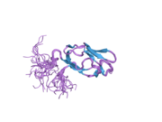 2dnc: Solution Structure of RSGI RUH-054, a lipoyl domain from human 2-oxoacid dehydrogenase
 2f5z: Crystal Structure of Human Dihydrolipoamide Dehydrogenase (E3) Complexed to the E3-Binding Domain of Human E3-Binding Protein
 2f60: Crystal Structure of the Dihydrolipoamide Dehydrogenase (E3)-Binding Domain of Human E3-Binding Protein


