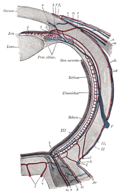
Ora serrata
| Ora serrata | |
|---|---|
 Diagram of the blood vessels of the eye, as seen in a horizontal section. | |
| Identifiers | |
| TA98 | A15.2.04.006 |
| TA2 | 6780 |
| FMA | 58600 |
| Anatomical terminology [edit on Wikidata] | |
The ora serrata is the serrated junction between the choroid and the ciliary body. This junction marks the transition from the simple, non-photosensitive area of the ciliary body to the complex, multi-layered, photosensitive region of the retina. The pigmented layer is continuous over choroid, ciliary body and iris while the nervous layer terminates just before the ciliary body. This point is the ora serrata. In this region the pigmented epithelium of the retina transitions into the outer pigmented epithelium of the ciliary body and the inner portion of the retina transitions into the non-pigmented epithelium of the cilia. In animals in which the region does not have a serrated appearance, it is called the ora ciliaris retinae.
Additional images
-
 Interior of anterior half of bulb of eye.
 -
 Vessels of the choroid, ciliary processes, and iris of a child.
 -
 Partial section of the human eye
See also
- Human eye
External links
- Anatomy photo:29:22-0204 at the SUNY Downstate Medical Center
- Atlas image: eye_1 at the University of Michigan Health System - "Sagittal Section Through the Eyeball"
- Atlas image: eye_3 at the University of Michigan Health System - "Coronal Section Through the Eyeball"
- "Anatomy diagram: 02566.000-1". Roche Lexicon - illustrated navigator. Elsevier. Archived from the original on 2012-07-22.
- v
- t
- e
(outer)
| Sclera |
|
|---|---|
| Cornea |
|

tunic (middle)
| Choroid | |
|---|---|
| Ciliary body | |
| Iris |
| Layers | |
|---|---|
| Cells |
|
| Other |
|
of the eye
| Anterior segment | |
|---|---|
| Posterior segment |
- Keratocytes
- Ocular immune system
- Optical coherence tomography
- Eye care professional
- Eye disease
- Refractive error
- Accommodation
- Physiological Optics
- Visual perception
 | This article about the eye is a stub. You can help Wikipedia by expanding it. |
- v
- t
- e


