60S ribosomal protein L26

Protein found in humans
RPL26
Available structures
PDBOrtholog search: PDBe RCSB
List of PDB id codes

4UG0, 4V6X, 5AJ0, 3J92, 4UJC, 3J7P, 4UJE, 3J7Q, 3J7R, 4D67, 4UJD, 4V5Z, 4D5Y, 3J7O

Identifiers
AliasesRPL26, DBA11, L26, ribosomal protein L26
External IDsOMIM: 603704; MGI: 106022; HomoloGene: 113207; GeneCards: RPL26; OMA:RPL26 - orthologs
Gene location (Human)
Chromosome 17 (human)
Chr.Chromosome 17 (human)[1]
Chromosome 17 (human)
Genomic location for RPL26
Band17p13.1Start8,377,516 bp[1]
End8,383,213 bp[1]
Gene location (Mouse)
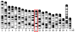
Chromosome 11 (mouse)
Chr.Chromosome 11 (mouse)[2]
Chromosome 11 (mouse)
Genomic location for RPL26
Band11|11 B3Start68,792,409 bp[2]
End68,797,815 bp[2]
RNA expression pattern
Bgee
HumanMouse (ortholog)
Top expressed in
  • Achilles tendon

  • ganglionic eminence

  • left ovary

  • right ovary

  • granulocyte

  • monocyte

  • lymph node

  • corpus callosum

  • canal of the cervix

  • uterine tube
Top expressed in
  • medial ganglionic eminence

  • transitional epithelium of urinary bladder

  • hair follicle

  • efferent ductule

  • migratory enteric neural crest cell

  • dermis

  • endothelial cell of lymphatic vessel

  • abdominal wall

  • skin of external ear

  • fossa
More reference expression data
BioGPS
n/a
Gene ontology
Molecular function
  • protein binding
  • RNA binding
  • structural constituent of ribosome
  • mRNA 5'-UTR binding
Cellular component
  • cytosol
  • ribosome
  • membrane
  • large ribosomal subunit
  • intracellular anatomical structure
  • cytosolic large ribosomal subunit
  • extracellular exosome
  • nucleoplasm
  • nucleolus
  • cytoplasm
  • cytosolic ribosome
  • ribonucleoprotein complex
Biological process
  • protein biosynthesis
  • viral transcription
  • SRP-dependent cotranslational protein targeting to membrane
  • ribosomal large subunit biogenesis
  • translational initiation
  • nuclear-transcribed mRNA catabolic process, nonsense-mediated decay
  • rRNA processing
  • cytoplasmic translation
  • DNA damage response, signal transduction by p53 class mediator resulting in cell cycle arrest
  • cellular response to UV
  • positive regulation of translation
  • cellular response to ionizing radiation
  • cellular response to gamma radiation
  • positive regulation of DNA damage response, signal transduction by p53 class mediator resulting in transcription of p21 class mediator
  • positive regulation of intrinsic apoptotic signaling pathway in response to DNA damage by p53 class mediator
  • regulation of translation involved in cellular response to UV
Sources:Amigo / QuickGO
Orthologs
SpeciesHumanMouse
Entrez

6154

19941

Ensembl

ENSG00000161970

ENSMUSG00000060938

UniProt

P61254

P61255

RefSeq (mRNA)

NM_000987
NM_001315530
NM_001315531

NM_009080

RefSeq (protein)

NP_000978
NP_001302459
NP_001302460

NP_033106

Location (UCSC)Chr 17: 8.38 – 8.38 MbChr 11: 68.79 – 68.8 Mb
PubMed search[3][4]
Wikidata
View/Edit HumanView/Edit Mouse

60S ribosomal protein L26 is a protein that in humans is encoded by the RPL26 gene.[5][6]

Function

Ribosomes, the organelles that catalyze protein synthesis, consist of a small 40S subunit and a large 60S subunit. Together these subunits are composed of 4 RNA species and approximately 80 structurally distinct proteins. This gene encodes a ribosomal protein that is a component of the 60S subunit. The protein belongs to the L24P family of ribosomal proteins. It is located in the cytoplasm. As is typical for genes encoding ribosomal proteins, there are multiple processed pseudogenes of this gene dispersed through the genome and the human paralog RPL26L1.[6]

Interactions

RPL26 has been shown to interact with Mdm2.[7]

See also

References

  1. ^ a b c GRCh38: Ensembl release 89: ENSG00000161970 – Ensembl, May 2017
  2. ^ a b c GRCm38: Ensembl release 89: ENSMUSG00000060938 – Ensembl, May 2017
  3. ^ "Human PubMed Reference:". National Center for Biotechnology Information, U.S. National Library of Medicine.
  4. ^ "Mouse PubMed Reference:". National Center for Biotechnology Information, U.S. National Library of Medicine.
  5. ^ Zaman GJ (May 1993). "Sequence of a cDNA encoding human ribosomal protein L26 and of a cDNA probably encoding human ribosomal protein L6". Nucleic Acids Res. 21 (7): 1673. doi:10.1093/nar/21.7.1673. PMC 309386. PMID 8479925.
  6. ^ a b "Entrez Gene: RPL26 ribosomal protein L26".
  7. ^ Ofir-Rosenfeld Y, Boggs K, Michael D, Kastan MB, Oren M (Oct 2008). "Mdm2 regulates p53 mRNA translation through inhibitory interactions with ribosomal protein L26". Mol. Cell. 32 (2): 180–9. doi:10.1016/j.molcel.2008.08.031. PMC 2587494. PMID 18951086.

Further reading

  • Wool IG, Chan YL, Glück A (1996). "Structure and evolution of mammalian ribosomal proteins". Biochem. Cell Biol. 73 (11–12): 933–47. doi:10.1139/o95-101. PMID 8722009.
  • Kenmochi N, Kawaguchi T, Rozen S, Davis E, Goodman N, Hudson TJ, Tanaka T, Page DC (1998). "A map of 75 human ribosomal protein genes". Genome Res. 8 (5): 509–23. doi:10.1101/gr.8.5.509. PMID 9582194.
  • Uechi T, Tanaka T, Kenmochi N (2001). "A complete map of the human ribosomal protein genes: assignment of 80 genes to the cytogenetic map and implications for human disorders". Genomics. 72 (3): 223–30. doi:10.1006/geno.2000.6470. PMID 11401437.
  • Andersen JS, Lyon CE, Fox AH, Leung AK, Lam YW, Steen H, Mann M, Lamond AI (2002). "Directed proteomic analysis of the human nucleolus". Curr. Biol. 12 (1): 1–11. Bibcode:2002CBio...12....1A. doi:10.1016/S0960-9822(01)00650-9. PMID 11790298. S2CID 14132033.
  • Yoshihama M, Uechi T, Asakawa S, Kawasaki K, Kato S, Higa S, Maeda N, Minoshima S, Tanaka T, Shimizu N, Kenmochi N (2002). "The Human Ribosomal Protein Genes: Sequencing and Comparative Analysis of 73 Genes". Genome Res. 12 (3): 379–90. doi:10.1101/gr.214202. PMC 155282. PMID 11875025.
  • Ewing RM, Chu P, Elisma F, Li H, Taylor P, Climie S, McBroom-Cerajewski L, Robinson MD, O'Connor L, Li M, Taylor R, Dharsee M, Ho Y, Heilbut A, Moore L, Zhang S, Ornatsky O, Bukhman YV, Ethier M, Sheng Y, Vasilescu J, Abu-Farha M, Lambert JP, Duewel HS, Stewart II, Kuehl B, Hogue K, Colwill K, Gladwish K, Muskat B, Kinach R, Adams SL, Moran MF, Morin GB, Topaloglou T, Figeys D (2007). "Large-scale mapping of human protein–protein interactions by mass spectrometry". Mol. Syst. Biol. 3 (1): 89. doi:10.1038/msb4100134. PMC 1847948. PMID 17353931.

External links

  • PDBe-KB provides an overview of all the structure information available in the PDB for Human 60S ribosomal protein L26


  • v
  • t
  • e
Proteins
Initiation factor
Bacterial
Mitochondrial
Archaeal
  • aIF1
  • aIF2
  • aIF5
  • aIF6
Eukaryotic
eIF1
eIF2
eIF3
eIF4
eIF5
eIF6
Elongation factor
Bacterial/​Mitochondrial
Archaeal/​Eukaryotic
Release factor
Ribosomal Proteins
Cytoplasmic
60S subunit
40S subunit
Mitochondrial
39S subunit
28S subunit
Other concepts
Stub icon

This protein-related article is a stub. You can help Wikipedia by expanding it.

  • v
  • t
  • e